Dense Breasts, Mammogram
Dense Breasts Explained: Why Some Women May Need More Than a Mammogram
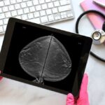
Dense breasts have more glandular and fibrous tissue than fat. On a mammogram, dense tissue shows up white and so do most tumours so small cancers can be harder to spot. Women with dense breasts may benefit from additional imaging, such as breast ultrasound or 3D mammography (digital breast tomosynthesis), alongside their routine mammogram. Breast density can only be confirmed by a mammogram, not by how breasts look or feel.
Breast density describes the proportion of glandular and fibrous (connective) tissue compared with fatty tissue, as seen on a mammogram. Radiologists grade it using four BI-RADS categories, from almost entirely fatty (A) to extremely dense (D). Categories C and D are considered “dense.”
Density has nothing to do with breast size or firmness – two women with similar-looking breasts can have very different density readings. It is also common: studies have found that Asian women tend to have a higher prevalence of dense breast tissue than Caucasian women, even after accounting for age and body weight.
| BI-RADS category | Description | Considered dense? |
|---|---|---|
| A | Almost entirely fatty | No |
| B | Scattered areas of density | No |
| C | Heterogeneously dense | Yes |
| D | Extremely dense | Yes |
Density matters for two separate reasons.
This combination is harder to detect and higher baseline risk is why density is worth understanding rather than ignoring.
You cannot tell by touch, and neither can your doctor by examination alone. Breast density is determined only when a radiologist reads your mammogram, and in many reports it is recorded as a BI-RADS density category. If you have had a mammogram, you can ask your doctor what your density category was. As a general pattern, breast tissue tends to become less dense with age as more fatty tissue develops, which is one reason mammograms often become easier to interpret in older women.
These are complementary tools, not competing ones. A doctor recommends the right combination based on your age, density, symptoms and personal risk.
| Method | How it works | Where it helps | Things to note |
|---|---|---|---|
| Mammogram (2D) | Low-dose X-ray of the breast | The recommended first-line screening test; the only method with proven mortality reduction in population screening (MOH / Singapore Cancer Society) | Sensitivity is reduced in dense breasts |
| 3D mammography (digital breast tomosynthesis, DBT) | Takes multiple thin X-ray “slices” to build a pseudo-3D image | May improve detection in dense breasts and can reduce unnecessary call-backs | Still an X-ray; availability and cost vary by clinic |
| Breast ultrasound | Uses sound waves; often used as a supplement | Can pick up some cancers hidden on a mammogram in dense breasts (studies report a few additional cancers detected per 1,000 women) | No radiation, but tends to produce more false positives |
| Breast MRI | Magnetic imaging | Usually reserved for women at high risk (e.g. strong family history, genetic factors) | Not a routine screening test for average-risk women |
The takeaway: for women with dense breasts, the question is often not “which one test,” but whether a single mammogram is enough on its own — and that is a conversation to have with a doctor.
Singapore’s national guidance (Health Promotion Board, Ministry of Health and Singapore Cancer Society) provides a useful baseline:
If your mammogram report shows dense breasts, that is worth raising with your doctor to decide whether supplementary imaging is appropriate for you. Fusion Medical offers digital breast tomosynthesis (3D mammography) and can advise on supplementary breast ultrasound where suitable, as part of its broader women’s health screening services.
Dense breasts are common and not a disease. They do, however, make mammograms harder to read and are linked to a modestly higher breast cancer risk, so they are a reason to be a little more thorough about screening — not a reason to panic.
No. Density refers to tissue composition on a mammogram, not firmness or size, so it can only be determined from imaging.
Having dense breasts does not mean you will get breast cancer, but it is associated with a slightly higher risk compared to less dense breast tissue.
Possibly. Supplementary ultrasound can detect some cancers missed by mammography in dense breasts, but it also produces more false alarms. Whether it is right for you depends on your overall risk — a doctor can advise.
Mammograms are generally recommended every two years from age 50, and from age 40–49. Women at higher risk may be advised to start earlier.
The compression is similar to a standard mammogram. Some women find it comparable; any discomfort is usually brief.
This article is for general information and is not a substitute for personalised medical advice. Please consult a doctor about your individual screening needs.
In Singapore, about 430,000 adults aged 18–69 have pre-diabetes, and without changes to diet and activity, roughly one in three goes on to develop type 2 diabetes within eight years.
Learn about lung cancer in Singapore, including key statistics, symptoms, causes in non-smokers, screening options, and why early detection matters.

Cancer screening in Singapore is no longer just age-based. Learn which screenings matter, who should consider them, and why early detection helps.

Dr Wenus Ho is a family physician and a designated workplace doctor with more than a decade of clinical experience. She is also currently a postgraduate tutor for the Graduate Diploma of Family Medicine (GDFM) Training Program in the Yong Yoo Lin School of Medicine, Singapore....

After graduating from Guys’, King’s and St Thomas’ School of Medicine in the United Kingdom, Dr Juliana went on to complete her Graduate Diploma in Family medicine at the National University of Singapore. She also has completed her diploma in Occupational Medicine. Dr Juliana applies her...

Dr Yong graduated from University College London, United Kingdom, with a Bachelor of Medicine & Bachelor of Surgery with distinction in medical sciences. She has more than 10 years of experience as a general practitioner, and was accredited as family physician by the Family Physician Accreditation...
Start your skin health restoration journey with us today!

501 Orchard Rd #04-11, Singapore 238880

Mon-Fri :8:30am – 5:30pm
Sat :8:30am – 1:00pm
Sun / PH :Closed
1 Fusionopolis Link, #01-05 Nexus
@ One-North , Singapore 138542
Mon-Fri :8:30am – 5:30pm
Sat/Sun / PH :Closed


Fat Freeze + X Wave at S$98
Vanquish ME + X Wave at S$199

