

Several images in one sweep
While the breast is compressed, the X-ray tube moves through a short arc and records several low-dose images. A 3D mammogram takes a little longer than a 2D one, but it feels much the same.



3D mammogram · Singapore
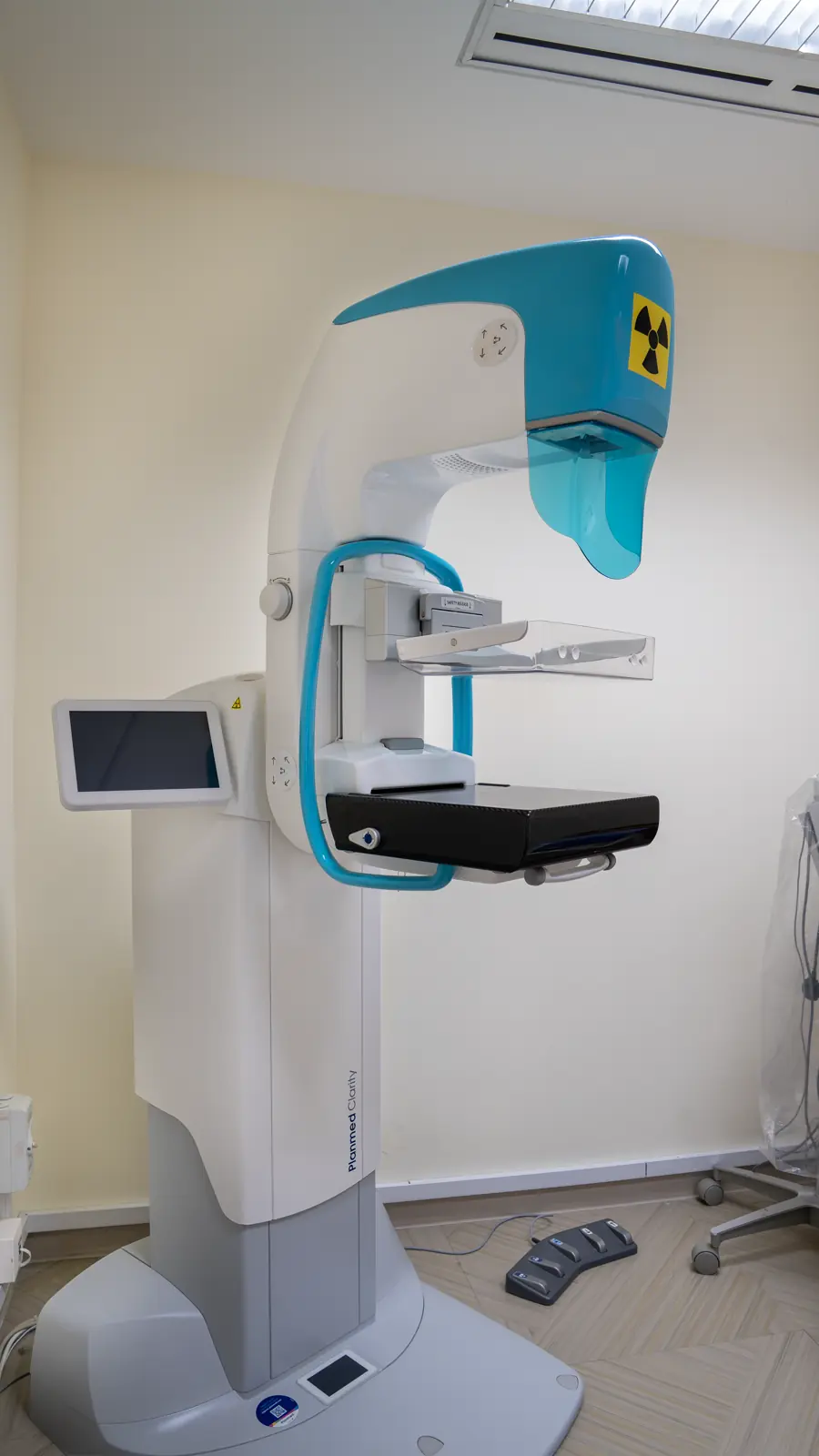
A 3D mammogram takes low-dose X-rays from several angles and builds them into thin slices, so breast tissue can be checked layer by layer. It is done on site at Wheelock Place.
A mammogram that shows the breast layer by layer, not as one flat picture.
A 3D mammogram, or digital breast tomosynthesis (DBT), is a low-dose X-ray of the breast taken from several angles. A computer rebuilds the images into thin slices, so the breast can be examined one layer at a time instead of as a single flat view. This can make overlapping or dense tissue easier to read.
At Fusion Medical, 3D mammograms are done on site at Wheelock Place, and 2D and 3D images can be taken in one sitting. A 3D mammogram is included in the Perimenopause & Menopause package and is one of the choices in Pearl (Female) and Breast & Women's Health. In Deluxe, Gold, Diamond and Opal (Female), you can switch to a 3D mammogram for a top-up.
How it works
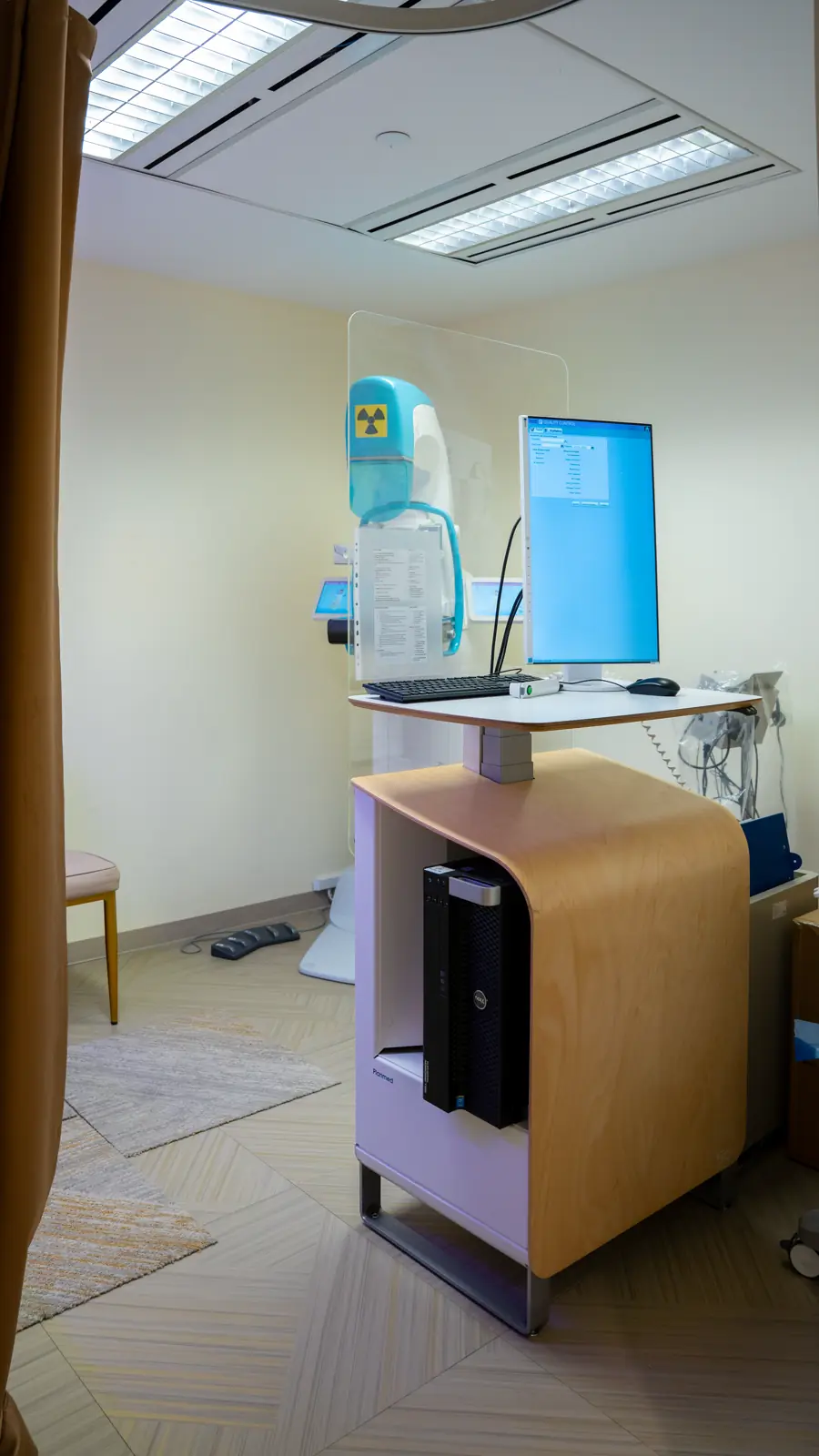
Each breast is positioned and compressed for a few seconds, as for a standard mammogram. The X-ray tube then moves in a short arc, taking several low-dose images that a computer rebuilds into thin slices. Looking through them one layer at a time reduces the effect of overlapping tissue.
Several images in one sweep
While the breast is compressed, the X-ray tube moves through a short arc and records several low-dose images. A 3D mammogram takes a little longer than a 2D one, but it feels much the same.

When your report says 'dense'
Breast density shows only on a mammogram, and your report may record it. Dense tissue and cancers both look white on a 2D image, so thin slices can help the radiologist see past the overlap. No test removes every limit of dense tissue.
When an ultrasound is added
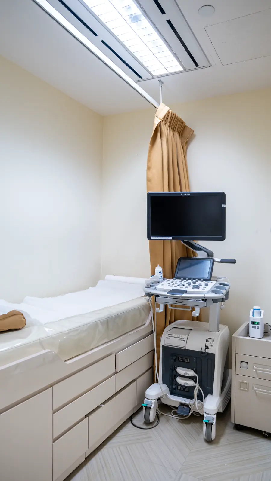
A breast ultrasound uses sound waves, not X-rays. It is often added to check a lump or an area seen on the mammogram, and your doctor may suggest it when breasts are dense.
Who it's for
A 3D mammogram can be used for screening or to look more closely at an area, depending on the question your doctor is asking. It can be particularly helpful when breast tissue is dense or an area is hard to assess on a 2D image. Your doctor may still suggest an ultrasound, extra views or another test.
New lump, nipple change or skin change? See a doctor now; don't wait for screening.
Choose 3D images for your screening mammogram after talking it through with your doctor.
Thin slices can help the radiologist see past overlapping dense tissue.
A recall or previous finding needs a plan from your doctor; 3D views may be part of it.
The Perimenopause & Menopause package includes a 3D mammogram.
The Breast & Women's Health package includes a 3D mammogram or a breast ultrasound.

Your 3D mammogram, step by step
Choose a date about a week after your period, and tell us about pregnancy, breastfeeding, implants or any breast symptoms.
Skip deodorant, powder and lotion on the day, wear a two-piece outfit and bring previous images and reports.
A female radiographer positions each breast in turn. The unit records the required 2D and 3D images while the breast is compressed for a few seconds.
The images are reported, and your doctor explains the results and arranges an ultrasound or further tests if needed.
Inside the clinic
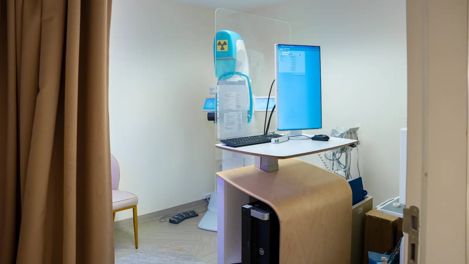
Our co-founders, Dr Juliana and Dr Wenus, start their tour of the clinic in the mammography room, where both 2D and 3D mammograms are done.
Safety & expectations
A 3D mammogram shows more layers, not every answer. A new breast change still needs a doctor, even after a normal result.
Tell us before your test if you are pregnant or breastfeeding, have implants or fillers, or have had breast surgery, and bring previous imaging. Keep checking your breasts, and see a doctor about any new change.
Fusion Medical performs 3D mammograms (digital breast tomosynthesis) on site at Wheelock Place, 501 Orchard Road, and 2D and 3D images can be taken in the same sitting. A 3D mammogram is included in, or can be added to, several of our health screening packages, and Singapore Citizens and PRs aged 50 and above can use MediSave for it. Our team can advise which option suits you, and our doctors can go through the results with you.
Not quite. Both use low-dose X-rays and the same kind of breast compression. A standard 2D mammogram takes flat pictures of each breast, while a 3D mammogram adds images from several angles that are rebuilt into thin slices, so the breast can be looked at layer by layer. Read more in 2D vs 3D mammogram: what is the difference?
A 3D mammogram (digital breast tomosynthesis) can help if you have dense breasts, because thin image slices reduce the tissue overlap that can hide an abnormality on a standard 2D mammogram. It does not remove every limitation of dense tissue, so a normal result cannot rule out every problem. Depending on your overall risk, your doctor and radiologist may still suggest an ultrasound or other imaging.
A 3D mammogram can be uncomfortable, but the compression is similar to a standard 2D mammogram and lasts only a few seconds for each view. Booking about a week after your period, when breasts are less tender, can help. Tell the radiographer if it hurts, as your position can often be adjusted.
If you still have periods, book about a week after your period. On the day, don't apply deodorant, perfume, powder, lotion or ointment to your breasts or underarms, as they can show up on the images. Wear a two-piece outfit, and bring previous mammograms and reports so any change can be compared.
Breast implants do not usually rule out a mammogram, but tell the clinic when you book about any implants or injected breast fillers, so it can confirm whether 3D images suit you. Implants can hide some breast tissue on X-ray, so extra views are often taken with the tissue eased forward. In breast screening, only gentle pressure is used, and there is currently no evidence that this causes implant rupture.
Tell the clinic before booking. A mammogram uses X-rays, and pregnancy or breastfeeding can change whether, when and how breast imaging is done. A breast ultrasound, which uses no radiation, is often the first test for a breast lump during pregnancy.
No. An abnormal result means an area needs a closer look, and most recalls turn out not to be cancer. Extra mammogram views, an ultrasound, an MRI or a small tissue sample (biopsy) may be recommended. Your doctor explains each step and what the results mean for you.
Appointments
Our team can tell you which packages include a 3D mammogram and help you choose between 2D and 3D imaging.